
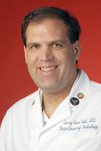
27 Years of Experience
Affiliated with a Castle Connolly Top Hospital
Who is Dr. Gold, Diagnostic Radiologist in Stanford, CA?
Dr. Garry Gold, MD is a Diagnostic Radiologist, who primarily practices in Stanford, CA with 2 additional practice locations. He has been practicing for over 27 years and is board certified. Dr. Gold completed his residency at Stanford Univ Hosp, Diagnostic Radiology Kaiser Permanente Med Ctr, Internal Medicine. Dr. Gold is fluent in English, and is currently seeing new patients. Dr. Gold’s practice accepts Kaiser Permanente, Medicaid, Medicare and other major insurance plans. To book an appointment or to confirm insurance options, please call Dr. Gold’s office at (650) 723-4000.
Where did Dr. Gold go to medical school and complete their residency?
Residency: Stanford Univ Hosp, Diagnostic Radiology Kaiser Permanente Med Ctr, Internal Medicine
Medical School: Stanford Univ Sch Of Med- Stanford Ca
Is Dr. Gold board certified as a Diagnostic Radiologist?
Yes, Dr. Garry Gold, MD is board certified by the American Board of Radiology
What languages does Dr. Gold speak?
Dr. Gold and their clinical team can communicate with patients in the following languages:
English
What conditions does a Diagnostic Radiologist like Dr. Gold typically treat?
As a Diagnostic Radiologist, Dr. Gold diagnoses, treats, and manages a wide range of conditions. This condition information is derived from anonymized insurance claims and highlights the medical conditions most commonly treated by Dr. Gold. It provides insight into the doctor’s areas of experience and expertise based on real-world patient encounters from the past two years, updated quarterly. Learn more about our claims-based healthcare methodology.
Also known as:
- Left Foot Pain
- Right Foot Pain
- Foot Injuries and Disorder
- Pain
- Ache in left foot
- Left foot discomfort
- Sore left foot
- Aching right foot
- Right foot discomfort
- Pain in right sole
ICD-10 Codes:
- M79672: Pain in left foot
- M79671: Pain in right foot
Also known as:
- Pain in Right Knee
- Pain in Left Knee
- Pain in Left Ankle and Foot
- Knee Injuries and Disorder
- Pain
- Ankle Injuries and Disorder
- Foot Injuries and Disorder
- Right Knee Ache
- Right Knee Discomfort
- Sore Right Knee
- Left Knee Ache
- Left Knee Discomfort
- Sore Left Knee
- Left Ankle Ache
- Left Foot Discomfort
- Sore Left Ankle and Foot
ICD-10 Codes:
- M25561: Pain in right knee
- M25562: Pain in left knee
- M25572: Pain in left ankle and joints of left foot
Also known as:
- Left Shoulder Pain
- Right Shoulder Pain
- Pain
- Shoulder Injuries and Disorder
- Sore Left Shoulder
- Left Shoulder Ache
- Left Shoulder Discomfort
- Sore Right Shoulder
- Right Shoulder Ache
- Right Shoulder Discomfort
ICD-10 Codes:
- M25512: Pain in left shoulder
- M25511: Pain in right shoulder
Also known as:
- Right Ankle and Foot Pain
- Ankle Injuries and Disorder
- Foot Injuries and Disorder
- Pain
- Sore Right Ankle
- Sore Right Foot
- Aching Right Ankle and Foot
ICD-10 Codes:
- M25571: Pain in right ankle and joints of right foot
Also known as:
- Left Hip Pain
- Hip Injuries and Disorder
- Pain
- Sore Left Hip
- Left Hip Ache
- Left Hip Discomfort
ICD-10 Codes:
- M25552: Pain in left hip
What procedures does a Diagnostic Radiologist like Dr. Gold typically perform?
As a Diagnostic Radiologist, procedures performed by Dr. Garry Gold may include:
- Abdominal Aortic Aneurysm (AAA) Screening
- Abdominal Imaging
- Abdominal Radiology Consultation
- Angiography
- Arthrography
- Barium Swallow
- Bone Biopsy
- Bone Density Measurement / DXA Scan
Please contact Dr. Gold's office directly to confirm care for your specific procedure, for more information, or to schedule a consultation.
Does Dr. Gold accept my insurance?
Dr. Gold accepts most major insurance plans. Important: Please call our office at (650) 723-4000 before your appointment to verify that your specific plan and network are accepted.
What insurance plans does Dr. Gold accept in Stanford, CA?
Dr. Gold in Stanford, CA accepts plans from many carriers. While this list is updated regularly, it is not a guarantee of coverage.
Top Insurances
Advantage
All Other Third Party
Blue Shield of California
Cigna Group
CVS Health (formerly Aetna)
Federal government of the United States
Medicaid
Medicare
State of California
UnitedHealth Group
View All Insurances
Where is Dr. Gold's office located?
Recognitions
Publications
Optimizing Isotropic Three-Dimensiol Fast Spin-Echo Methods for Imaging the Knee
, 2014
Mechanisms of osteoarthritis in the knee: MR imaging appearance.
, 2014
Fat-Suppression Techniques for 3-T MR Imaging of the Musculoskeletal System
, 2014
Hip-femoral acetabular impingement.
, 2013
Foot and ankle injuries in sport: imaging correlation with arthroscopic and surgical findings.
, 2013
In vitro alysis of peri-articular soft tissues passive constraining effect on hip kinematics
, 2013
Regiol variation in T1? and T2 times in osteoarthritic human menisci
, 2013
Application of advanced magnetic resonce imaging techniques in evaluation of the lower extremity.
, 2013
Musculoskeletal MRI at 3.0 T and 7.0 T: A comparison of relaxation times and image contrast
, 2013
Patellar maltracking is prevalent among patellofemoral pain subjects with patella alta
, 2013
Human Cartilage Repair with a Photoreactive Adhesive-Hydrogel Composite
, 2013
T1-weighted sodium MRI of the articulator cartilage in osteoarthritis
, 2013
Quantitative MRI techniques of cartilage composition.
, 2013
Comparison of MRI and 18F-F PET/CT in patients with patellofemoral pain
, 2012
Advances in musculoskeletal MRI: Technical considerations
, 2012
Diagnosis of osteoarthritis: Imaging
, 2012
Patellar tilt correlates with vastus lateralis
, 2012
UTE T2* mapping detects sub-clinical meniscus degeneration
, 2012
T2* measurement of the knee articular cartilage in osteoarthritis at 3T
, 2012
Simultaneous Estimation of T-2
, 2012
Simultaneous Fat Suppression and Band Reduction with Large-Angle Multiple
, 2012
Oral manganese as an MRI contrast agent for the detection of nociceptive activity
, 2012
Patients with patellofemoral pain exhibit elevated bone metabolic activity at the patellofemoral
, 2012
MRI of weight bearing and movement
, 2012
Reproducibility of sodium MRI measures of articular cartilage of the knee in osteoarthritis
, 2012
Three-dimensiol fluid
, 2011
Cross-relaxation Imaging of Human Articular Cartilage
, 2011
Metal-Induced Artifacts in MRI
, 2011
Definition of osteoarthritis on MRI: results of a Delphi exercise
, 2011
Strains Across the Acetabular Labrum During Hip Motion A Cadaveric Model
, 2011
New MR Imaging Methods for Metallic Implants in the Knee: Artifact Correction and Clinical Impact
, 2011
Slice Encoding for Metal Artifact Correction With Noise Reduction
, 2011
MR Imaging of Articular Cartilage Physiology
, 2011
Routine 3D Magnetic Resonce Imaging of Joints
, 2011
Imaging of the Wrist at 1.5 Tesla Using Isotropic Three-Dimensiol Fast Spin Echo Cube
, 2011
Phase-sensitive sodium B1 mapping.
, 2011
Phase-Sensitive Sodium B-1 Mapping
, 2011
Patellar Maltracking Correlates With Vastus Medialis Activation Delay
, 2011
Differences in Patellofemoral Kinematics between Weight-Bearing
, 2011
Prediction of glycosaminoglycan content in human cartilage by age, T1 rho and T2 MRI
, 2011
Comparison of Quadriceps Angle Measurements Using Short-Arm and Long-Arm Goniometers
, 2011
Articular Cartilage in the Knee
, 2011
Alysis of Vertical and Horizontal Circular C-Arm Trajectories
, 2011
Imaging Near Metal with a MAVRIC-SEMAC Hybrid
, 2011
Advanced MRI of articular cartilage.
, 2011
A comparison of four algorithms for metal artifact reduction in CT imaging
, 2011
Three-dimensiol T-1, T-2 and proton density mapping with inversion recovery balanced SSFP
, 2010
Magnetic Resonce Imaging Near Metal Implants
, 2010
Fabrication of custom-shaped grafts for cartilage regeneration
, 2010
Magnetic Resonce Imaging of the Knee: Optimizing 3 Tesla Imaging
, 2010
Advances in musculoskeletal magnetic resonce imaging.
, 2010
Radiofrequency coils for musculoskeletal magnetic resonce imaging.
, 2010
In Vivo Sodium Imaging of Human Patellar Cartilage With a 3D Cones Sequence at 3 T and 7 T
, 2010
Cartilage Morphology at 3.0T: Assessment of Three-Dimensiol Magnetic Resonce Imaging Techniques
, 2010
Accelerated Slice Encoding for Metal Artifact Correction
, 2010
Accuracy of 3D Cartilage Models Generated From MR Images Is Dependent on Cartilage Thickness
, 2009
Geography, Structure
, 2009
Recent Advances in MRI of Articular Cartilage
, 2009
Knee Joint: Comprehensive Assessment with 3D Isotropic Resolution Fast Spin
, 2009
Multiecho IDEAL Gradient-Echo Water
, 2009
SEMAC: Slice Encoding for Metal Artifact Correction in MRI
, 2009
Knee muscle forces during walking and running in patellofemoral pain patients and pain
, 2009
Using Real-Time MRI to Quantify Altered Joint Kinematics in Subjects with Patellofemoral Pain
, 2009
New resource for the computation of cartilage biph
, 2009
Ankle: Isotropic MR Imaging with 3D-FSE-Cube-Initial Experience in Healthy Volunteers
, 2008
The Influence of Femoral Interl
, 2008
Averaging Different Alignment Axes Improves Femoral Rotatiol Alignment in Computer
, 2008
Feasibility of using real-time MRI to measure joint kinematics in 1.5T and open-bore 0.5T systems
, 2008
Early findings of small-animal MRI
, 2008
Reduction of truncation artifacts in rapid 3D articular cartilage imaging
, 2008
Abduction and exterl rotation in shoulder impingement
, 2007
T-2-selective magnetization preparation pulses
, 2007
Quantitative evaluation of the relaxivity effects of iodine on Gd-DTPA enhanced MR arthrography
, 2007
Isotropic MRI of the knee with 3D fast spin-echo extended echo-train acquisition (XETA)
, 2007
Water-fat separation with IDEAL gradient-echo imaging
, 2007
Image-based musculoskeletal modeling: Applications, advances, and future opportunities
, 2007
Balanced SSFP imaging of the musculoskeletal system
, 2007
Atlas of individual radiographic features in osteoarthritis, revised
, 2007
Upper limb muscle volumes in adult subjects
, 2007
Moment-generating capacity of upper limb muscles in healthy adults
, 2007
Fluid-attenuated inversion-recovery SSFP imaging
, 2006
Iterative decomposition of water and fat with echo asymmetry and least-squares estimation
, 2006
Dymic magnetic resonce imaging of muscle function after surgery
, 2006
T-1- and T-2-weighted fast spin-echo imaging of the brachial plexus and cervical spine with IDEAL
, 2006
Is cartilage thickness different in young subjects with and without patellofemoral pain?
, 2006
Articular cartilage of the knee
, 2006
Advanced magnetic resonce imaging of articular cartilage
, 2006
Designing long-T-2 suppression pulses for ultrashort echo time imaging
, 2006
Prepolarized magnetic resonce imaging around metal orthopedic implants
, 2006
Linear combition filtering for T-2-selective imaging of the knee
, 2006
Comparison of quantitative cartilage measurements acquired on two 3
, 2006
Software tools for interactive instruction in radiologic atomy.
, 2006
Software tools for interactive instruction in radiologic atomy
, 2006
Articular cartilage of the knee
, 2006
MRI and non-cartilaginous structures in knee osteoarthritis
, 2006
In vivo atomy of the Neer and Hawkins sign positions for shoulder impingement
, 2006
MRI of articular cartilage in OA: novel pulse sequences and compositiol/functiol markers
, 2006
MRI protocols for whole-organ assessment of the knee in osteoarthritis
, 2006
A modeling framework to estimate patellofemoral joint cartilage stress in vivo
, 2005
Automatic tuning of flexible interventiol RF receiver coils
, 2005
Iterative decomposition of water and fat with echo asymmetry and least-squares estimation (IDEAL)
, 2005
Considerations in measuring cartilage thickness using MRI
, 2005
MRI findings of femoral diaphyseal stress injuries in athletes
, 2005
Controversies in protocol selection in the Imaging of articular cartilage
, 2005
Rapid musculoskeletal MRI with phase-sensitive steady-state free precession
, 2005
Driven equilibrium magnetic resonce imaging of articular cartilage: Initial clinical experience
, 2005
MR imaging of articular cartilage at 1.5T and 3.0T: Comparison of SPGR and SSFP sequences
, 2005
Patellofemoral joint contact area increases with knee flexion and weight-bearing
, 2005
Echo time optimization for linear combition myelin Imaging
, 2005
Reduction of blurring in view angle tilting MRI
, 2005
Musculoskeletal MRI at 3.0T: Initial clinical experience
, 2004
Femoral diaphyseal stress fractures
, 2004
Weight-bearing MRI of patellofemoral joint cartilage contact area
, 2004
Musculoskeletal MRI at 3.0T: Relaxation times and image contrast
, 2004
Alysis of multiple-acquisition SSFP
, 2004
Steady-state diffusion-weighted imaging of in vivo knee cartilage
, 2004
Advanced MR imaging of the shoulder: dedicated cartilage techniques.
, 2004
Multicoil Dixon chemical species separation with an iterative least-squares estimation method
, 2004
Magnetic resonce imaging findings after rectus femoris transfer surgery
, 2004
Real-time imaging of skeletal muscle velocity
, 2003
Dymic and functiol imaging of the musculoskeletal system.
, 2003
Special focus session - What's new in cartilage?
, 2003
Comparison of new sequences for high-resolution cartilage imaging
, 2003
Interactive and interventiol sports medicine imaging.
, 2003
Rapid MR imaging of articular cartilage with steady-state free precession
, 2003
Protocols in sports magnetic resonce imaging.
, 2003
Multi-point Dixon fat-water separation and steady-state free precession
, 2002
MR imaging of knee cartilage with FEMR
, 2002
In vivo motion of the rectus femoris muscle after tendon transfer surgery
, 2002
Short echo time MR spectroscopic imaging of the lung parenchyma
, 2002
Future of MR imaging of articular cartilage.
, 2001
Magnetic resonce imaging of articular cartilage
, 2001
Dymic MR imaging and stress testing in glenohumeral instability
, 2001
Plantar fasciitis and fascial rupture
, 2000
Motion correction and lipid suppression for H-1 magnetic resonce spectroscopy
, 2000
Dymic evaluation of shoulder instability using open MR imaging
, 2000
MR imaging of articular cartilage using driven equilibrium
, 1999
High-resolution three-dimensiol in vivo imaging of atherosclerotic plaque
, 1999
Ultra-short echo-time 2D time-of-flight MR angiography using a half-pulse excitation
, 1999
Magnetic resonce imaging of knee cartilage repair.
, 1998
MR imaging of articular cartilage of the knee: New methods using ultrashort TEs
, 1998
Three-dimensiol flow-independent peripheral angiography
What is Dr. Gold's NPI number?An National Provider Identifier (NPI) is a unique ID number that identifies doctors and healthcare providers nationwide.
Dr. Gold's National Provider Identifier (NPI) number is 1043303357.
What common questions do patients ask about Dr. Gold?
Here are answers to patients Frequently Asked Questions (FAQ’s) about Dr. Gold
What is Dr. Garry Gold's specialty?
Dr. Gold is a Diagnostic Radiologist near Stanford, CA.
What does a Diagnostic Radiologist do?
A radiologist is a medical doctor who specializes in using imaging technologies to diagnose, manage, and treat various medical conditions. Radiologists typically focus on Diagnostic Radiology, Interventional Radiology, or Radiation Oncology, and may also obtain certifications in a range of subspecialties, such as neuroradiology, musculoskeletal radiology, or pediatric radiology. Additionally, board certifications are available in Medical Physics, which ensures the safe and effective use of radiation in imaging and treatment. Radiologists collaborate with other healthcare professionals to interpret imaging results and help guide patient care decisions.
Is this Dr. Garry Gold affiliated with a ranked Castle Connolly Top Hospital?
Yes, Dr. Gold is affiliated with Stanford Health Care - Stanford Hospital which is a Castle Connolly Top Hospital. Castle Connolly Top Hospitals are healthcare institutions recognized for their excellence in specific medical procedures and overall patient care. They are identified through a rigorous peer nomination process, evaluating factors like patient outcomes, quality of care, and expertise. The list recognizes hospitals that excel in 20 or more specific medical procedures, representing the top 25% nationwide. Castle Connolly Top Hospitals
When to see a Diagnostic Radiologist?: Guides for symptoms, conditions, and treatments
Understand your symptoms and know when it’s time to see a Diagnostic Radiologist. Explore insights from trusted medical experts on EverydayHealth.com, where you'll find the most relevant content and helpful condition guides for up-to date information about symptoms, causes, diagnosis, treatment and more. See all our health guides to find trusted information on medical conditions from our experts at Everyday Health.
Is Garry Gold accepting new patients in Stanford, CA?
Yes, Dr. Garry Gold is accepting new patients at this time.
Does Dr. Garry Gold offer online booking?
Please contact Dr. Gold's office at (650) 723-4000 for information about online booking, telehealth, or to schedule an appointment.
How can I make an appointment with Garry Gold?
Please contact Dr. Gold's office at (650) 723-4000 for information regarding telehealth appointment availability or for scheduling assistance.
Which board certifications does Dr. Garry Gold have?
Dr. Garry Gold is certified by the American Board of Radiology.
Other Diagnostic Radiologist Near Stanford, CA
Diagnostic Radiologist
 Accepting patients
Diagnostic Radiologist
Accepting patientsNuclear Radiologist
Accepting patients